Skin Conditions Around The Eyes
Dr Christophe Hsu – dermatologist. Geneva, Switzerland
Eyelid Contact Dermatitis
- Cosmetics and cleansers are usually used on the eyelids or eyelashes. These can cause an irritant or allergic reaction in patients sensitive to such cosmetics and cleansers.
- Patients will complain of itching, burning and redness.
- Signs of contact dermatitis include blistering, redness and scaliness.
- If an allergy is suspected a patch test can be done to confirm an allergy and to ascertain the causative cosmetic or chemical responsible. Avoidance of the cosmetic will cure the condition.
- Eye drops, contact lens and medicaments may also cause eyelid contact dermatitis. If you have eyelid dermatitis you should consult your doctor who will prescribe some creams and refer you to a dermatologist for allergy test if necessary.
Atopic Dermatitis
- Atopic Dermatitis is a genetic skin disorder.
- Patients with atopic dermatitis often have eyelid involvement. The skin may be red, scaly and sometimes oozing. Itch is common.
- Sometimes the eye lining and eyelids are also affected in atopic dermatitis and a sore eye may result. There is watery eye discharge and patients may find their eyes sensitive to light. If the eyelid or eye itself is affected your doctor will prescribe some eyedrops and creams for you. Creams will help.
- Many patients are also allergic to housedust. Housedust may aggravate their eye inflammation·
Bacterial Skin Infection (Impetigo or Conjunctivitis)
- Like skin elsewhere, the eyelids can be infected by bacteria.
- The eye will show a thick, sticky yellow discharge and the eyelids may be red with yellow crusts.
- Impetigo is most commonly seen in children.
- Good hygiene and antibiotics are needed to clear the bacterial infection.
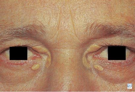
Xanthelasma
- These are flat to slightly raised yellowish plaques on the upper and lower eyelids.
- They are associated with high blood cholesterol or triglycerides level in about 20% of people with this condition. Sometimes there will be a family history of similar problem.
- The xanthelasma can be excised surgically or by laser or chemical treatment.
- Such lesions may recur.
- Patients with high cholesterol or triglyceride should have their lipid level controlled before surgery.

Xanthelasma
Syringoma
- These are tiny skin coloured growths on the eyelids.
- Several family members may have the same lesions.
- These growths are harmless.
- They are enlarged growths, under developed sweat glands.
- Syringomas usually appear during adolescent and adulthood.
- Most people choose to leave these skin growths alone. These can be removed surgically by laser or excision for cosmetic reason.
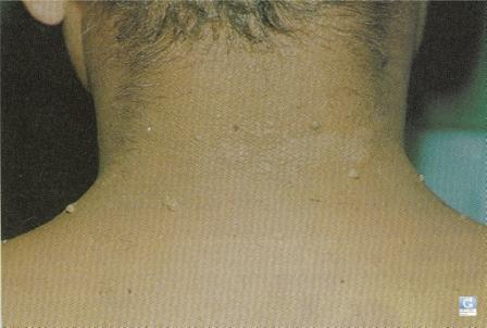
Skin Tags (Acrochordon)
- Skin Tags Small outgrowths of skin can be seen around the eyes and the eyelids in some individuals.
- Similar skin outgrowths are often seen on the neck and chest.
- These growths are skin coloured and harmless.
- Treatment is not necessary but they can be easily removed surgically for cosmetic reason.

Skin tags
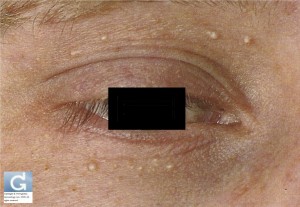
Milia
- These are small white or yellowish white skin growths often seen on the eyelids or temple.
- They are very small and resemble millet seeds.
- They can be removed surgically for cosmetic reasons.
- They represent obstructed sweat ducts.
Milia
Dark Rings Round The Eyes
- Darkened pigmentation of the eyelids are common in many dark skin individuals.
- In many cases the darkening seems to vary with stress or lack of sleep.
- This condition is benign and may be inherited or constitutional.
- It is not a sign of any illness.
- Treatment is unnecessary. There is no effective treatment against the condition.
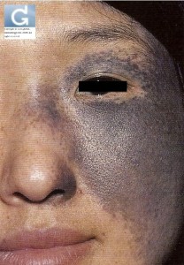
Naevus of Ota
- This is a birthmark that occurs at birth or shortly after birth as a patch of blue black discolouration on one cheek, temple and eyelids.
- Usually one side of the face is affected although occasionally both sides of the {ace may be blue, black discolouration is present on the white of the eye.
- The pigmentation of Naevus of Ota can be reduced by pigment laser surgery. Patients will require multiple treatments at 2-3 monthly interval. Only the pigmentation on the skin can be treated.
Naevus of Ota
Strawberry Naevus
- These are vascular birthmarks.
- It can appear as a large red soft growth on the eyelids.
- The growth will continue to enlarge and grow as the baby grows old but the growth will slowly regress spontaneously when the child is 3-4 years old:
- If the vascular growths are small, they can be left alone to await spontaneous regression.
- If large and encroaching on the eyelid, vision may be affected. They should then receive treatment. Treatment options include oral medication, injections and laser ablation.
Portwine Stain
- There is another vascular birthmark.
- They present as flat red patches on the eyelids and the skin of the cheek at birth.
- Unlike strawberry naevus, portwine stains never disappear spontaneously as the child grows older:
- The vascular birthmarks increase in thickness and small blebs of blood vessels may be seen.
- Agsin, these growths do not regress on their own and treatment is necessary. Most portwine stains respond to the vascular laser. Patients will need multiple laser treatments at 3 monthly intervals to achieve an optimal result.
Contributors
Dr Christophe Hsu – dermatologist. Geneva, Switzerland
National Skin Centre. Singapore
العربية 中文-汉语 中文-漢語 Deutsch Español Italiano 日本語 Português 한국어 русский язык Tagalog
Category : atopic dermatitis - Modifie le 11.29.2009Category : cernes - Modifie le 11.29.2009Category : contact dermatitis - Modifie le 11.29.2009Category : darkening around the eyes - Modifie le 11.29.2009Category : dermatite atopique - Modifie le 11.29.2009Category : Eczéma de contact - Modifie le 11.29.2009Category : excroissances - Modifie le 11.29.2009Category : infections - Modifie le 11.29.2009Category : kystes miliaires - Modifie le 11.29.2009Category : milia - Modifie le 11.29.2009Category : névus d'Ota - Modifie le 11.29.2009Category : nevus fraise - Modifie le 11.29.2009Category : nevus of Ota - Modifie le 11.29.2009Category : skin growths - Modifie le 11.29.2009Category : strawberry nevus - Modifie le 11.29.2009Category : syringoma - Modifie le 11.29.2009Category : syringome - Modifie le 11.29.2009Category : xanthélasma - Modifie le 11.29.2009Category : xanthélasme - Modifie le 11.29.2009


