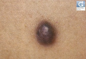
Dermatofibroma
Dr Christophe HSU – dermatologist. Geneva, Switzerland
What is dermatofibroma ?
- It is a benign condition
- It typically appears in females 9 times more often than in males.
- It is also known as superficial benign fibrous histiocytoma.
What is it caused by ?
- The exact cause of its development is unknown.
- There was an inital theory that it was a reaction following an insect bite. It is also hypothesized to be a neoplastic growth.
- Hypothesis: The female predominance and its occurence in young age suggests that hormonal factors may play a role.
What does it look like ?
- It presents as dermal bumps which are typically present on the legs.
- It displays the dimple sign when compressed laterally: the lesion becomes depressed.
- The nodules can be dark-coloured. This is because of the deposit of hemosiderin. (In those cases, it can mimick malignant melanoma)
When examined by a dermatologist with a dermoscopy, the most typical pattern is a stellate white central pattern surrounded by a rim of hyperpigmentation. At the surface, a pigmented network may be present.
Is it painful ?
- The lesions are most often asymptomatic
- On occasion, they can itch and/or be painful
Is it a cause for concern ?
- Isolated lesions carry no significant burden. They are often a cause for concern and can be mistaken for other benign (moles) or malignant conditions.
- Numerous lesions may be the marker of systemic diseases such as Systemic Lupus Erythematosus.
- Associations have been described for HIV infection. However, dermatomyositis, Graves disease, Hashimoto thyroiditis, myasthenia gravis, Down syndrome, leukemia, myelodysplastic syndrome, cutaneous T-cell lymphoma, multiple myeloma, and atopic dermatitis have all been reported in association with the phenomenon. In addition, antiretroviral agents and the biologic agent efalizumab have been linked to their appearance.
The dermatologist might perform a biopsy in case of diagnosis doubt and /or order other tests.
How to treat ?
- Treatment is warranted for cosmetic reasons and when symptoms are present.
- Excision is a possibility. However because of its frequent location on the legs, unsightly scars can be a consequence.
- Cryotherapy may be an option before excision but there is a risk of post-inflammatory hyperpigmentation.
Contributors:
Dr Christophe HSU – dermatologist. Geneva, Switzerland
Italiano 日本語 Deutsch русский язык
Category : Dermatofibrome - Modifie le 11.6.2012Category : Histiocytofibrome - Modifie le 11.6.2012


