Introduction (For Professionals)
Seborrheic keratosis (SK) is a benign epidermal neoplasm which presents as an eruption of one to hundreds of papules or plaques. The major potential burden for the patient is a cosmetic nuisance, but a florid acute appearance of SK lesions can rarely be associated with an internal neoplasm {Vielhauer, 2000; Heaphy, 2000; Grob, 1991}. Sometimes, SK may be found together with basal cell carcinomas or spinal cell carcinomas and can be confused with malignant melanoma. The epidemiologically very different dermatosis papulosa nigra is sometimes considered as a variant of seborrheic keratosis as well as the differently distributed stucco keratosis (Braun falco, 1978) as a hypopigmented variant of seborrheic keratosis. These two latter conditions will not be developed in this review.
Historical Features
SK was first described clinically and histologically by Frudenthal in 1926 {Frudenthal, 1926}. SK was already suspected as a separate entity earlier than Freudenthal since Holländer related them to internal cancer. SK was already considered as a seperate entity by Neumann (1832-1906){Crissey, 2002} Again, during the Third International Congress of Dermatology held in London in 1898, Dubreuilh debated the aetiology of “varieties of keratosis” with P. Unna and Brooke {Dubreuilh, 1898}. Dubreuilh emphasized that a separate kind of lesion, subsequently named SK, had to be distinguished from actinic keratosis and warts, and that it did not evolve into skin cancer.
Epidemiology
SK is the most prevalent benign tumor of the skin. It usually appears under the age of forty and progresses in number intraindividually and interindividually.
The developpment of SK does not seem correlated to sex, hair color or eye color. In a study of 170 individuals in a population aged 15 to 30 {Gill, 2000}, there was no significant difference between the two sexes, nor any link between the presence of SK and eye or hair color.
Prevalence does seem to vary with the gender according to Young (1965). In an analysis of 222 patients in a home ward, 37,9% of women compared to 29,3% of men had SK{Young, 1965}. Under the age of forty this difference is more marked with 8,3% of males and 16,7% of females in the UK presenting with at least one lesion {Memon, 1995}.
According to Yeatman and al {Yeatman, 1997} , SK affects an increasing number of people with advancing age and a greater number of lesions for every affected individual is formed. In this study, 100 individuals were classified into age groups : 12% of individuals aged 15 to 25 presented a SK (23% of individuals between 15 inad 30 in another study{Gill, 2000}) , whereas each patient over 75 was reported to have SK. This result can be compared with 88% of patients over 64 having at least one SK, according to another study {Tindall, 1963}. According to a study in korean males{Kwon, 2003}, the greatest increase in prevalence of individuals with a seborrheic keratosis is between ages 40 and 50, where the increase in prevalence increases from 78.9% to 93.9%. At 60 years of age the prevalence reaches 98.7%.
In the same study{Yeatman, 1997} the average number of lesions for one patient increases in average from 6 in the 15-25 age group to 69 in patients aged over 75 with an intermediary 23 lesions in patients aged 51 to 75.
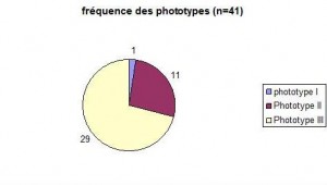
SK is present in younger people in countries with more UV (B and C) exposure. Yeatman (1997) compares the prevalence of SK in his study group of patients under the age of forty in Australia with a comparable group in the United Kingdom {Memon, 1995}. He found that 18,2% of males and 32% of females in the Australian group group had SK. Another way of quatifying the potential role of sunlight is objecting the greater number of lesions on photoexposed skin compared with non-exposed skin {Kwon, 2003}. At age 40, that ratio is of 5.7 , 11.2 at 50 and 18.3 at age 60. There was however no statistical significance between the decreased prevalence of SK from Fitzpatrick’s phototypes I-III compared with Fitpatrick’s phototypes IV-VI. In our experience, in a study of 41 SKs, light phototypes didn’t seem more affected than darker phototypes. Over 70% of patients with SK had a type III phototype. Nevertheless, we do not know the prevalence of phototypes in Geneva, Switzerland. This small group does did not include brown skin (phototype IV).
Histologically, certain types are found more frequently than others. Maize and coll. {Maize, 1995} studied 108 SK prospectively. 71 (66%) of lesions were acanthotic, 27 (25%) hyperkeratotic, 10 (9% reticulated). None of the lesions was of the clonal type. Neither pigmentation nor irritation were taken into account (verifier).
SK, as a whole, has been shown to be present at 15 years of age. On the other hand, inverted follicular keratosis (IFK) doesn’t appear before age 50.
This advice is for informational purposes only and does not replace therapeutic judgement done by a skin doctor.
Contributors:
Dr Christophe HSU – dermatologist. Geneva, Switzerland
Category : épidémiologie - Modifie le 10.12.2012Category : historical facts - Modifie le 10.12.2012Category : historique - Modifie le 10.12.2012Category : introduction - Modifie le 10.12.2012Category : kératose séborréhique - Modifie le 10.12.2012Category : seborrheic keratoses - Modifie le 10.12.2012Category : Seborrhoeic keratosis - Modifie le 10.12.2012Category : verrue séborrhéique - Modifie le 10.12.2012


